Unveiling The Hidden Wonders: A Deep Dive Into Sponges Under The Microscope
At first glance, a sponge might seem like a simple, unassuming organism. Often mistaken for plants due to their sessile nature, these ancient creatures are, in fact, fascinating animals. They lack true tissues and organs, yet possess an incredible complexity that truly comes to life when viewed through the powerful lens of a microscope. Stepping into the microscopic world of a sponge is like discovering a hidden city, bustling with intricate structures and specialized cells working in harmony. What wonders await us when we put a sponge under the microscope?
The Architectural Marvels: Spicules, the Sponge's Skeleton
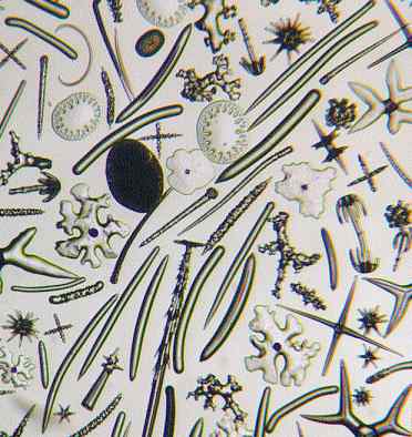
If sponges are the buildings of the marine world, then their spicules are undoubtedly the "bricks." These incredible structures are the primary components of a sponge's skeleton, providing both structural support and a unique defense mechanism. Without them, a sponge would simply crumble into an amorphous blob, which is precisely what happens when a spicule-rich sponge dies and its soft tissues decay, leaving behind only these sparkling, needle-like remnants.
Composition and Classification of Spicules
Sponge spicules are made of one of two primary materials:
- Calcium Carbonate: These spicules are characteristic of sponges belonging to the phylum Calcarea. They are often less intricate in shape compared to their silica counterparts.
- Silica: Composed of silicon dioxide, these spicules are found in sponges of the phylum Silicea. They are renowned for their often fantastic and elaborate shapes.
The size of spicules also varies significantly. Large spicules, often visible to the naked eye, are referred to as megascleres (or macroscleres). However, the true artistry of sponge architecture lies in the smaller, microscopic ones, aptly termed microscleres. These tiny structures require a powerful compound microscope to fully appreciate their intricate designs.
Unique Shapes and Functions
One of the most remarkable aspects of spicules is their diversity. The shapes, sizes, and overall composition are absolutely unique for each species of sponge. From simple rods and needles to complex stars, anchors, and grappling hooks, these microscopic crystals are truly wonders of natural engineering. Together, these features are crucial for species identification, making spicules invaluable tools for marine biologists and paleontologists.
Beyond providing the essential framework that gives the sponge its characteristic shape, spicules also serve as a formidable defense against predators. Imagine trying to eat a creature made of tiny, sharp glass or crystal needles – not a pleasant meal!
- Alicia Lyons
- 1500 Calorie Meal Plan Indian Non Veg
- Traditional Tattoo Artist
- Brown British Short Hair Cat
- Barriga Com
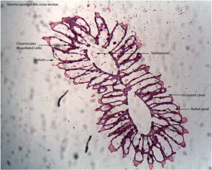
The Inner Workings: Channels, Pores, and Choanocytes
While spicules provide the skeleton, the true lifeblood of a sponge flows through its intricate internal channel system. Sponges are filter feeders, and their entire existence revolves around moving water through their bodies to extract food and oxygen, and to expel waste. This complex plumbing system is a marvel of biological efficiency.
The Role of Choanocytes
Central to this water flow system are specialized cells called choanocytes, or "collar cells." These remarkable cells line the internal channels of the sponge and possess a single, whip-like appendage known as a flagellum. The synchronized beating of countless flagella creates a powerful current that draws water into the sponge through numerous tiny openings called pores (ostia).
Once inside, the water courses through the internal channels, and the choanocytes capture microscopic food particles with their collar-like structures. This efficient system allows sponges to bring in vital food and oxygen, and simultaneously to take out carbon dioxide and other metabolic waste products. The filtered water then exits the sponge through a larger opening, typically located at the top, called the osculum. Examining these structures under a compound microscope provides a fascinating view of this continuous, life-sustaining flow.
Survival and Reproduction: The Gemmule
Sponges are incredibly resilient creatures, capable of surviving harsh environmental conditions. One of their most remarkable adaptations for survival and asexual reproduction is the formation of a gemmule. These structures are essentially "survival capsules" that allow sponges to endure periods of drought, freezing, or lack of food, and then regenerate when conditions improve.
Under the microscope, a sponge gemmule reveals its intricate design:
- Inner Protoplasmic Part: This core contains undifferentiated cells, essentially the "building blocks" ready to develop into a new sponge.
- Membranous Coats: Surrounding the protoplasmic part are one or more protective membranous layers, offering initial protection.
- Pneumatic Coat or Granular Crust: Outside of these inner membranes, there is usually a more robust outer layer, often described as a pneumatic coat (filled with air pockets for buoyancy or insulation) or a granular crust, providing significant protection against environmental stressors.
These miniature fortresses are a testament to the sponge's evolutionary success, ensuring the continuation of the species even when faced with extreme adversity.
Beyond the Biological: The Microbial World in Sponges
While our primary focus has been on the biological structures of marine sponges, it's worth noting that any sponge-like material, whether natural or synthetic, can harbor a thriving microscopic ecosystem. For instance, a used dishwashing sponge, when placed under a microscope, can reveal an astonishing bacterial world. From 40x to 400x magnification, one can witness countless microorganisms, demonstrating that even in our everyday objects, a hidden microscopic universe exists, teeming with life.
Why Explore Sponges Under a Microscope?
The act of examining a sponge under a microscope is more than just a scientific exercise; it's an eye-opening journey into the hidden complexity of seemingly simple life forms. Here's why it's so valuable:
- Unveiling Biodiversity: The unique shapes and compositions of spicules for each species highlight the incredible diversity within the sponge phyla, Calcarea and Silicea.
- Understanding Fundamental Biology: Observing choanocytes in action, or the structure of a gemmule, provides direct insight into basic biological processes like filter feeding, water circulation, and asexual reproduction.
- Educational Value: For students, seeing the "whole sponge animal" and then delving into the "structures inside the sponge’s pores" through a compound microscope offers a comprehensive and engaging view of these fascinating creatures, making abstract concepts tangible.
- Appreciation for Nature: It fosters a deeper appreciation for the intricate designs and survival strategies present even in organisms that might appear unremarkable at first glance.
Conclusion
From the sparkling, structurally vital spicules that form their unique skeletons and provide defense, to the ceaselessly beating flagella of choanocytes driving water through intricate internal channels for feeding and respiration, and the resilient gemmules ensuring survival, sponges are truly microscopic marvels. Exploring these ancient animals under a microscope reveals a world of sophisticated biological engineering and adaptation, reminding us that even the simplest-looking organisms hold a universe of complexity waiting to be discovered. It’s a journey that transforms our understanding of life itself, one microscopic detail at a time.
Porifera - Dr. Peat's Biology Page
Sponge Spicules

BIO-102 LAB--SPONGE CROSS SECTION UNDER MICROSCOPE - YouTube
